Scientists Use AI and Synchroton Radiation CT Imaging to Improve Lithium Batteries
Widespread use of lithium-ion batteries in portable electronics and electric vehicles has made improving battery performance a key research focus in recent years. Since battery particle cracks seriously degrade lithium-ion battery performance, many scientists are focused on how to avoid such cracks. Recently, the 4W1A imaging experimental station of the Beijing Synchrotron Radiation Facility (BSRF), Institute of High Energy Physics, Chinese Academy of Sciences, cooperated with LIU Yijin's research group at the Stanford Synchrotron Radiation Light Source and Peter Cloetens’ research group at the European Synchrotron Radiation Facility (ESRF) to study the crack generation mechanism of commercial 18650 battery cathode material by using deep learning technology and synchrotron radiation, nano-resolution CT imaging technology.
The researchers first conducted three-dimensional imaging of tens of thousands of electrode particles in the cathode of a commercial 18650 battery through nano-resolution, X-ray CT imaging technology. They then proposed an image segmentation algorithm based on deep learning to quantitatively analyze the degree of damage to the battery. This algorithm overcomes the shortcomings of traditional image segmentation algorithms based on image grayscale and achieves automatic detection and extraction of crack information from tens of thousands of battery particles (Fig. 1). Finally, through quantitative statistics and analysis of massive battery cracks, the researchers studied the relationship between battery energy density and crack density (Fig. 2). These results indicate that, to reduce particle cracks, adaptive strategies of particle packing density for different depths should be considered. The above research will play an important role in guiding structural gradient design of electrode material in the depth direction and improvement of battery material performance. This study was published in Advanced Functional Materials. FU Tianyu (BSRF) and Federico Monaco (ESRF) are co-authors of the paper.
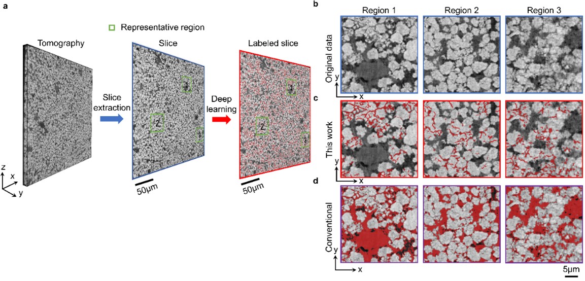
Fig. 1. Representative examples of crack detection results and comparison with a conventional approach. (a) The trained model is applied to each slice of tomography data and an overview of crack distribution is obtained. (b) Three selected regions. (c) and (d) Crack detection results obtained by our deep-learning method and by a conventional image-intensity-based segmentation approach, respectively.(Image by IHEP)
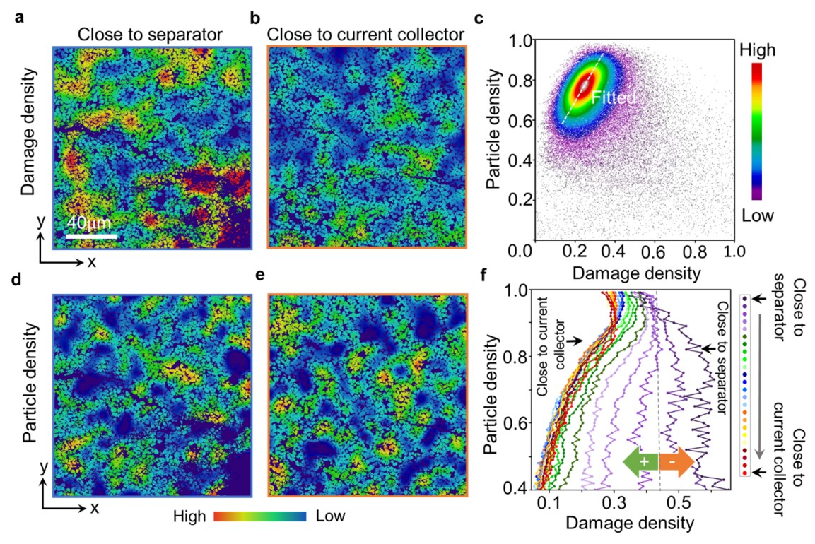
Fig. 2. Association of particle cracks with particle density. (a) and (c) Damage density map and particle density map of a slice close to the separator. (b) and (d) Damage density map and particle density map of a slice close to the current collector. (e) Correlation plot of particle density and damage density over the entire imaged 3D volume. The overall positive trend is indicated by the white fitted line. (f) Correlation between particle density and damage density is plotted layer-by-layer. A positive-to-negative transition is observed.(Image by IHEP)
Contact Information
Mr. GUO Lijun
ljguo@ihep.ac.cn
Related News

Multi-scale Synchroton X-ray Imaging Quantifies Li-ion Battery Failure
06-20,19
Researchers from the Beijing Synchroton Radiation Facility, the European Synchroton Radiation Fac...

Second Run of 2017 Concluded at BSRF
12-01,17
This year's second dedicated run at the Beijing Synchrotron Radiation Facility (BSRF) was open to...