How lasers cast a light on accelerator science (1)
Since their invention, lasers have played important roles in research at particle accelerators. This link is set to continue growing ever closer, as Chan Joshi explains.
Particle accelerators, invented during the first half of the 20th century, and lasers, invented during the latter half of the 20th century, are arguably the two most successful tools of scientific discovery ever
 |
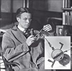
| Ernest Lawrence |
Lasers at light sources and accelerators
 |
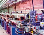
| Fig. 2 |
How are the high-brightness electron beams needed for X-ray FELs and for future colliders for high-energy particle physics produced? Here, again, lasers have played a critical role. Until the mid-1980s, thermionic cathodes embedded in RF cavities were used to produce electron bunches. It was realized that the emittance of beams from these RF guns could be greatly improved by replacing the thermionic cathode (LaB6 for instance) by a photocathode (Cu, Mg or alkali cathode). Richard Sheffield and colleagues at Los Alamos National Laboratory carried out pioneering work on the first photo-injector gun with a Cs3Sb cathode (Sheffield et al. 1996). By illuminating the photocathode with a short laser pulse of photons with an energy just greater than the work function of the cathode material and by operating the gun at high gradients, very high currents of electron bunches with emittances less than 1 mm mrad have been produced (Akre et al. 2008). For FELs, the short duration combined with low emittance implies a beam of high brightness that can be readily bunched by the FEL instability on the wavelength scale of the emitted radiation. Almost all recent FELs, including FLASH at DESY (FLASH: the king of VUV and soft X-rays), the high-gain harmonic-generation (HGHG) FEL at Brookhaven National Laboratory and the LCLS at SLAC, use photo-injector guns as the source of electrons.
Lasers are also routinely used for alignment and diagnostics in particle accelerators. A "laser wire" is a type of beam-profile monitor used to sample nondestructively the transverse profile of intense
 |
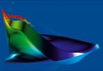
| Fig. 3 |
Future linear colliders for particle physics will bring nanometre-sized beams of electrons and positrons into head-on collision, maximizing the luminosity. Conventional techniques for measuring the size of the beam spot, such as the laser-wire scanning described above, are not suitable for measuring submicron spots. Instead, laser photon CS at 90° has been used as a noninvasive diagnostic technique for measuring ultrasmall beam sizes (Shintake 1992). Two counter-propagating laser beams produce a standing-wave interference pattern. As the focused electron bunch is scanned across this interference pattern (using a weak steering magnet) the gamma-ray yield is modulated and the depth of modulation of the CS gamma-ray flux gives the spot size. This technique has been used at the Final Focus Test Beam Facility at SLAC to measure a 47 GeV beam with a transverse size as small as 60 nm. A modified version of this technique using a propagating beat-wave interference pattern can be used as a bunch-length monitor.
Measurement of the width of highly relativistic electron bunches shorter than 100 fs poses a serious challenge. Fortunately, the transverse electric field of such bunches can itself be used to induce a change in the polarization of a synchronized laser pulse in an electro-optic crystal that is placed in the vicinity of the beam (Cavalieri et al. 2005). The duration of the photons that are affected by the polarization change can then be measured to give the pulse width of the electrons.
to be continued
Source: CERN COURIER
About the author
Chan Joshi, University of California Los Angeles.