Fast, safe and high-sensitive X-ray imaging, not far in the future
A research team from the Beijing Synchrotron Radiation Facility (BSRF), Institute of High Energy Physics made great
 |
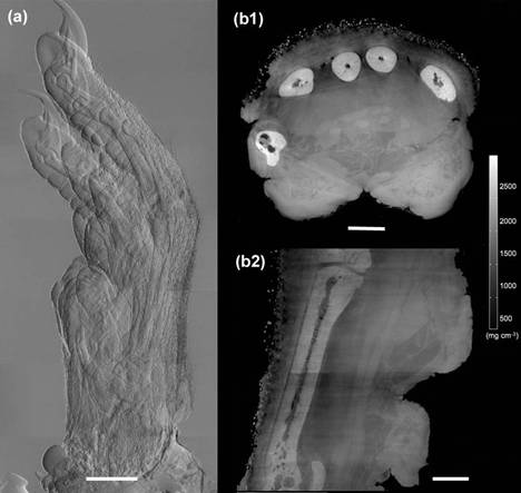
| Imaging of a rat paw. Structural details of hard tissue (bone), soft tissue (muscles, fat) and even hairs are well visible |
The scientific researchers presented an innovative, highly sensitive X-ray tomographic phase-contrast imaging approach. This new method, dubbed as "Reverse Projection" (RP) protocol, not only significantly reduced the delivered dose without the degradation of the image quality, but also gained much higher efficiency. The new technique set the prerequisites for fast and low-dose phase-contrast X-ray imaging methods in the future.
X-ray radiography and tomography were closely related to people’s daily life. The conventional absorption-based imaging was widely used in clinical diagnosis, biology, material science, information science and security check, etc. However, the X-ray-imaging community had been continually challenged by the frustrating question of how to increase the contrast in X-ray images with simple and fast methods and without increasing the dose imparted to a specimen.
According to the principle that the absorption contrast is the same but the refraction contrast opposite between the front projection and back projection, the research staff proposed RP protocol in hard X-ray phase contrast imaging to overcome some of the problems that have impaired the wider use of phase contrast in X-ray radiography and tomography.
In order to verify the feasibility of this proposal, the team conducted a series of scientific experiments with X-ray imaging experts from Tokyo University and Swiss Light Source. This RP protocol proved to significantly increase the sensitivity and phase contrast in soft tissue and polymeric materials. The paper titled “Low-dose, simple, and fast grating-based X-ray phase-contrast imaging” was published in PNAS, 107, 31, 13576-13581 (2010).
For the X-ray imaging community, who engaged in simplifying the method to extract the phase information in critical samples and minimizing delivery dose for a long time, this new approach meant a significant step forward. It is anticipated that implementation of the RP protocol in new medical X-ray CT scanners will produce fast, safe and high-sensitive X-ray imaging in the coming future.