New PET System for human body imaging developed
Scientists from the Institute of High Energy Physics (IHEP) recently developed a new type of high-resolution PET system for human body
 |
|
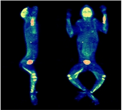
a sanned image of a 4.5kg macaque (image by IHEP) |
The new PET system has adopted technologies with self-owned intellectual properties, such as the detector, electronics, image reconstruction and domestic LYSO scintillation crystal. After the assembling and commissioning, the system went through a test of a body scanning of a macaque. The scanned image reflected a clear picture of its vivo metabolism. The image resolution of the cone field of view (CFOV) is less than 3.5mm, better than the standards of the National Electrical Manufacturers Association (NEMA). In the next phase, scientists will move to the registration and inspection for the medical facility and its clinical trials.
Over the years, IHEP has transformed its advantages in mega-science facilities, multi-disciplinary scientific research and the relevant high technologies into many high-tech products. In 1986, China’s first PET scanner for human body was developed. From 2006 to 2012, a new type of animal PET, dedicated PET scanner for breast imaging and animal PET/CT was developed, both being its first kind in China.
Positron Emission Tomography (PET) is a nuclear medical imaging technique that produces a three-dimensional image or picture of functional processes in the body. PET is both a medical and research tool. It is used heavily in clinical oncology (medical imaging of tumors and the search for metastases), and for clinical diagnosis of certain diffuse brain diseases such as those causing various types of dementias. PET is also an important research tool to map normal human brain and heart function, and to support drug development.