| Press |
| News |
| Upcoming Events |
| Reports from Overseas |
| Image Bank |
| IHEP Video |
 |
 |
 |
| Location: Home > Press > News |
| PEM System Developed by IHEP | TEXT SIZE: A A A | |
| —— | ||
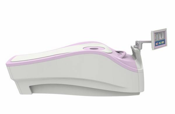
Positron emission mammography imaging system (PEM) is a breast-dedicated imaging system developed by the Institute of High Energy Physics (IHEP), the Chinese Academy of Sciences. Recently, PEM has got the Ⅲ category medical instrument registration license, which means the system is approved for sale and for clinical applications. The R&D efforts have been supported by "863" key research program under the Ministry of Science and Technology. To push technology transformation and PEM industrialization, IHEP has been in collaboration with Jingma Holding Group, Hangzhou Gaoneng Medical Equipment Ltd. The Clinical tests have been done in the Tumor Hospital of Tianjin Medical University and Xuanwu Hospital of Capital University of Medical Sciences. Clinical results show that PEM has great advantages in breast diagnosis. In addition, the efficacy and safety performances of PEM exceed original expectations. PEM has a number of advantages compared to whole-body PET, especially in the detection of early minute lesions, tumor location, differentiating benign or malignant tumors and adjuvant therapy evaluation. Positron emission tomography (PET) is a nuclear medicine technology, which is able to detect tumors' abnormal metabolism and to produce three-dimensional images of functional maps of the body. This kind of functional imaging techniques can detect tumors metabolism changes at an early stage. The PEM system is a breast-dedicated PET imaging system with high sensitivity and high specificity. Compared with other clinical anatomical imaging methods, such as mammography, ultrasound, CT, MRI, PEM helps achieve early breast cancer diagnosis by detecting metabolism changes of tumors. The PEM system adopts ring detector configuration. Patient just needs to lie prone with a pendant breast imaged uncompressed taking the examination. The 1.38 mm resolution performance could greatly improve the detection ability to small and early breast tumors. Breast cancer ranks the highest incidence rate and second mortality rate in female malignant tumors. The patient number is on the increase and the average age is becoming younger. Early detection, early diagnosis and early treatment are the keys to improve treatment effect, increase the survival rate and improve quality of life of the patients. The success of PEM not only upgrades the early diagnosis technology of breast cancer, but also promotes the capacity of independent innovation in high-end nuclear medical-imaging equipment field in China. (Registration number: 20153331166) (Performance test results and clinical application could be referred in “Performance Evaluation and Initial Clinical Test of the Positron Emission Mammography System, PEMi. IEEE Transactions on Nuclear Science”)
|
||||