| Cysteine–Ag Cluster Hydrogel Confirmed by Experimental and Numerical Studies |
| From: PublishDate:2016-06-03 Hits: |
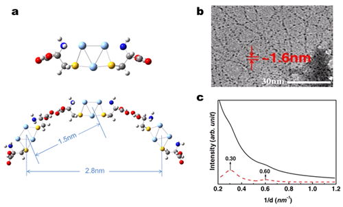
Noble metal clusters are composed of several to hundreds of atoms with ultrasmall size ( < 2 nm), including Ag3、Ag4、Au25 etc. The determinant chromophore can be tunable from ultraviolet to near-infrared fluorescence with high quantum yield. More important, the surface functionalization of cluster is promising for the cell targeting imaging. Very recently, the noble metal clusters have attracted much attention as a new material for cell imaging. The researchers from the nanoimaging and nanomedicine group (institute of high energy physics) have constructed a novel hydrogel system based on Ag3 cluster coated by the native cysteine (Cys) amino acid. They characterized the physicochemical properties and the formation mechanisms of the hydrogel by the combination method of experimental and numerical studies. The results have been published in small, 2015, 11 (38), 5118-25. The researchers have found three factors to ensure the self-assembly of Cys coating Ag3, and result in the hydrogel. First, the Ag–S bonds make Cys and Ag3 form Cys-Ag3-Cys monomer. Second, intermolecular hydrogen bonds between carboxyl groups of adjacent monomer push them self-assembled. Third, more monomers precisely self-assemble to produce the –[Cys-Ag3-Cys]n multimer, e.g., a single molecular chain with the left-handed helix conformation, via a benign thermodynamic process. These multimers entangle together to form micro-network to trap water and produce hydrogel in situ. For the biomedical application, the hydrogen bonds of hydrogel are sensitive to thermal and proton stimuli, and the hydrogel presents lysosome targeting properties via fluorescent imaging with biocompatibility. On the basis of the SAXS (BSRF, 1W2A), the researchers have studied the microstructure of the hydrogel. 2D SR-SAXS patterns of the hydrogel and blank Cys aqueous solution were investigated in details. The integrated SR-SAXS patterns of the hydrogel reveals two peaks in diagram c, with one at 0.3 (d ≈ 3.3 nm) and the other at 0.6 (d ≈ 1.7 nm). In the SR-SAXS pattern, the 3.3 nm spacing is comparable to the length between the first Ag3 and the third Ag3 in helix pitch of ≈ 2.8 nm from the theoretical simulation, and the 1.7 nm spacing corresponded to the spacing between adjacent Ag3 from the DFT calculation (1.5 nm) in diagram a and from HRTEM images (1.6 nm) along the backbone of [Cys-Ag3-Cys]n in diagram b. The small difference between the SR-SAXS/HRTEM and simulation results is acceptable when the experimental error is taken into consideration along with [Cys-Ag3-Cys]n multimer conformation relaxation in solution compared with the trimer in the numerical simulation. (http://onlinelibrary.wiley.com/wol1/doi/10.1002/smll.201501245) This study has provided an important clue to understand the process and mechanism of the hydrogel formation. In this research, the synchrotron light source (BSRF) helps to uncover the microstructure and the repeated unit information of the hydrogel, which provides a very good assistance for the design of cluster based hydrogel for the biomedical applications. Article: Yanyan Cui, Yaling Wang* and Lina Zhao*, Cysteine–Ag Cluster Hydrogel Confirmed by Experimental and Numerical Studies, Small 2015, 11 (38), 5118-25. |
|
|
| Chinese

News and Events
- Beamline 1W1 of BSRF started to runoperate in the couplingparasitic mode of BEPCII
- Synthesis of High Performance Polymer Materials for Field Effect-Transistors
- Surfactant molecular aggregates in green solvents
- GIXRD has played an important role in the characterization of organic thin-film transistors

Copyright © 2011 - 2012 Beijing Synchrotron Radiation Facility