
| Nanoprobes: Quantitatively Detecting the Femtogram Level of Arsenite Ions in Live Cells |
| From: PublishDate:2012-06-26 Hits: |
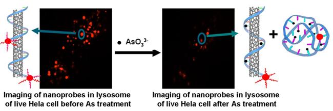
Recently, arsenite (As) have been applied to treat the acute promyelocytic leukemia (APL) successfully. Further research revealed that the apoptotic effects of arsenic are not restricted to APL cells but also can be observed in other malignant cells in vitro. There are at least three different molecular mechanisms are proposed but not uniform. Note that all these molecular mechanisms are contrary with each other and they all focused the pathway of bio-molecular triggered by As, no solid information of arsenite in live cell are supplied to match these molecular mechanisms, therefore it is hard for researchers to figure out the right molecular mechanism to understand how did arsenic kill cancer. A group from Institute of High Energy Physics designed a kind of fluorescent nanoprobes to high sensitivity detection the drug arsenic ions in live cells. The research has been published on June 2nd, 2011 in ACS nano. The team designed the fluorescent nanoprobe, single-strand DNA wrapped single-wall carbon nanotubes (ssDNA-SWCNTs, the ssDNA is labeled by the dye molecule), which can detect arsenite ions via an emission decrease method. The mechanism of the nanoprobes was studied by the SRCD in BSRF and AFM as follows. For the nanoprobe, the SWCNT is wrapped by ssDNA, and the ssDNA is labeled by the 50-hexachloro-fluorescein phosphoramidite (HEX). Arsenite ions could strongly bind the G/T bases of ssDNA and decrease the π-πinteraction between ssDNA and SWCNTs. This makes some ssDNA dissociate from the SWCNTs and further adapt condensed conformation in solution. Such condensed conformation of ssDNA will cause the HEX to interact with G/T base-binding arsenite ions, and further induce metal ions quenching the emission of HEX. Thus, the nanoprobes can detect arsenite ions via an emission decrease method. . With the help of a confocal microscope and cryo-electron microscopy, the lysosome target of the arsenite ion and nanoprobe is well described in high spatial resolution. In a live cell, trace arsenite ions could interact with nanoprobes and significantly decrease the emission of the nanoprobes. This result supported that high dosage arsenic will push the cancer cell apoptosis more quickly as more As is concentrated in the cell. For the first time, our studies provide a method to detect artificial metal ions via spatial and quantity parameters in live cells. This study also gives clear proof that the target organelle of arsenite is lysosome, and this strong proof can help the molecular researcher determine the right molecular pathway of As-induced cancer cell apoptosis.
Article: Ru Liu, Zhong Chen, Yaling Wang, Yanyan Cui, Huarui Zhu, Ping Huang, Wei Li, Yuliang Zhao*, Ye Tao, and Xueyun Gao*,Nanoprobes: Quantitatively Detecting the Femtogram Level of Arsenite Ions in Live Cells, ACS Nano, 2011, 5, 5560–5565. |
|
|
| Chinese
Science Highlights
Home /

Copyright © 2011 - 2012 Beijing Synchrotron Radiation Facility